Fluorescence In Situ Hybridization (FISH) is a pivotal molecular cytogenetic technique that enables the detection and localization of specific DNA sequences on chromosomes, facilitating precise genetic analysis in research and clinical settings.
Principles of FISH
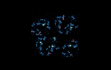
FISH involves the use of fluorescently labeled DNA or RNA probes that are complementary to sequences of interest. These probes are denatured and then hybridized to denatured chromosomal DNA or nuclei fixed on microscopic slides. Once binding (hybridization) occurs, the slides are washed to remove excess probes, and hybridized probes are visualized using fluorescence microscopy. The specific hybridization sites appear as bright signals, corresponding to the location of the targeted DNA sequences.
Applications in Cytogenetics
FISH has revolutionized cytogenetic analysis by offering unique advantages over traditional banding techniques:
- Detection of numerical and structural abnormalities: Enables identification of deletions, duplications, inversions, translocations, and aneuploidy in both metaphase and interphase cells.
- Oncology diagnostics: Used in the detection and characterization of chromosomal changes associated with hematological malignancies and solid tumors, even in samples unsuitable for conventional karyotyping such as formalin-fixed, paraffin-embedded tissues.
- Prenatal and postnatal diagnosis: Allows rapid assessment of chromosomal abnormalities in fetal cells obtained from amniocentesis or chorionic villus sampling.
- Gene mapping and genome organization: Facilitates assignment of genes to specific chromosomal locations.
- Detection of microdeletions and cryptic rearrangements: Identifies submicroscopic changes undetectable by standard karyotype analysis.
Key Advantages
Can be performed on non-dividing (interphase) cells, enabling analysis of samples otherwise inaccessible to standard cytogenetics.
Allows for use of multiple fluorescent probes simultaneously, providing detailed, multicolor imaging of several targets within a single cell.
High sensitivity and specificity for targeted sequences.
Fluorescence In Situ Hybridization remains a gold standard for targeted cytogenetic analysis, offering deep resolution, flexibility, and diagnostic value that continues to push the boundaries of genome research and personalized medicine.