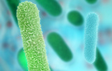
Special stains for microorganisms play a crucial role in microbiology and clinical pathology by providing enhanced visualization and differentiation of microbial pathogens in tissue or clinical samples that cannot be adequately distinguished by routine staining methods. These stains are indispensable for diagnosing infectious diseases and understanding microbial morphology and cellular structures.
Principle and Importance of Special Stains
Special stains exploit the unique chemical and structural properties of microorganisms to selectively color specific components such as cell walls, capsules, spores, or intracellular inclusions. By applying differential dyes or staining techniques, these methods reveal microorganisms that may be difficult to see with conventional stains like hematoxylin and eosin. They serve as complementary tools to immunohistochemistry, molecular diagnostics, and culture methods in clinical settings.
Applications in Medicine and Research
Special stains assist in rapid and accurate pathogen identification, enabling targeted antimicrobial therapy and improved patient outcomes. They are extensively used in histopathology to detect bacterial and fungal infections in biopsies, in parasitology for protozoan and helminth visualization, and in microbiological research to study microbial anatomy and pathogenic mechanisms.
Special stains for microorganisms remain essential diagnostic adjuncts in microbiology and pathology. By selectively highlighting microorganisms and their unique features, these stains enhance diagnostic precision and support clinical decision-making for infectious diseases. Mastery of special stain techniques is fundamental for microbiologists and pathologists alike, ensuring effective detection and understanding of microbial pathogens.