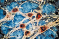
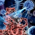
Flow cytometry is a powerful technique used for rapid multiparametric analysis of physical and chemical characteristics of individual cells or particles in suspension. It relies on fluorescently labeled antibodies to identify and quantify proteins and other cellular markers.
Overview of Flow Cytometry Protocol
1. Sample Preparation
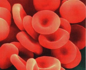
- Prepare a single-cell suspension from tissue, blood, or cultured cells using mechanical dissociation, enzymatic digestion, or gentle pipetting.
- For adherent cells, detach using enzymatic solutions or calcium chelators.
- Count and assess cell viability to ensure sufficient live cells for analysis.
2. Blocking and Staining
- Block nonspecific binding sites to reduce background.
- Incubate cells with fluorescently labeled antibodies for surface marker detection.
- For intracellular markers, fix and permeabilize cells after surface staining.
- Use viability dyes to distinguish live and dead cells.
3. Washing and Resuspension
- Wash cells thoroughly to remove unbound reagents.
- Resuspend cells in buffer compatible with flow cytometry.
4. Data Acquisition
- Run samples in a flow cytometer, where cells pass individually through laser beams.
- Fluorescence and light-scattering signals are detected to quantify cell markers and physical properties.
- Include controls for compensation and gating strategies.
5. Data Analysis
- Use specialized software to generate histograms and dot plots.
- Identify cell populations and quantify marker expression levels.
- Perform multiparametric analysis for detailed profiling.
Flow cytometry enables comprehensive immunophenotyping and functional characterization of cells in both research and clinical environments. Protocol adjustments may be necessary depending on sample type, target markers, and fluorophores used.