

Cryopreservation of Primary Human Dermal Fibroblasts

Table of contents
- 1. Introduction
- 2. Principle of cryopreservation
- 3. Cryopreservation Strategy
- 4. Equipment
- 5. Reagents and Consumables
- 6. Experimental Protocol
- 7. Preparation of Cryoprotective Medium
- 8. CPA Exposure and Cell Dispensing
- 9. Controlled Cooling
- 10. Cryogenic Storage
- 11. Warming and Post‑Thaw Recovery
- 12. CPA Dilution and Removal
- 13. Post-Thaw Culture
- 14. Quality Control Measures
- 15. Acceptance and Interpretation Criteria
- 16. Critical Parameters Requiring Laboratory Validation
- 17. Troubleshooting
- 18. Conclusion
- References
1. Introduction
Primary human dermal fibroblasts (HDFs) are adherent stromal cells isolated from human skin and are widely used in extracellular‑matrix biology, wound repair, tissue engineering, regenerative medicine, cellular reprogramming, and skin biology. Cryopreservation enables defined fibroblast populations to be stored for subsequent experimental use and reduces the need for continuous culture expansion.
Cryopreservation of primary fibroblasts is feasible, although post‑thaw recovery depends on cryoprotectant composition, cellular state, cooling conditions, storage history, and recovery procedures. This protocol describes the cryopreservation of primary HDFs as a cell suspension using controlled/gradual cooling and a dimethyl sulfoxide ( DMSO )‑containing cryoprotective medium.
The principal formulation described here is FBS + 10% DMSO (v/v) , which has been used for human fibroblast cryopreservation (Mohamed et al., 2024). Recent work has also investigated HES-containing, low- DMSO cryoprotective formulations for human dermal fibroblasts. For example, Zheng et al. (2024) evaluated HES with 2% DMSO as part of a low- DMSO cryopreservation strategy. This formulation should be treated as a separate experimental condition rather than as a modification of the 10% DMSO formulation.
2. Principle of cryopreservation
Cryopreservation of mammalian cells is governed by the interaction between extracellular ice formation, cellular water transport, solute concentration, membrane permeability, intracellular ice formation, and cryoprotectant toxicity (Whaley et al., 2021).
During cooling, extracellular water preferentially forms ice, progressively concentrating solutes in the remaining unfrozen fraction. This increases extracellular osmotic pressure and promotes movement of intracellular water toward the extracellular environment. The resulting cellular dehydration is an important component of controlled freezing (Whaley et al., 2021).
The cooling rate is therefore a critical variable. Controlled cooling allows sufficient water efflux and cellular dehydration before intracellular freezing becomes likely. Excessively rapid cooling may increase the probability of intracellular ice formation, whereas excessively slow cooling may produce excessive dehydration and prolonged exposure to concentrated extracellular solutes (Whaley et al., 2021).
DMSO is a penetrating cryoprotective agent that enters cells and modifies the physicochemical conditions associated with freezing. Its protective effect is associated with reduction of intracellular ice formation, while its toxicity depends on concentration, temperature, and exposure duration (Weng & Beauchesne, 2020).
Successful cryopreservation therefore requires an appropriate balance between cellular dehydration and protection against intracellular ice formation while limiting solution‑effect injury and cryoprotectant toxicity (Whaley et al., 2021; Weng & Beauchesne, 2020).
3. Cryopreservation Strategy
The procedure consists of the following sequential stages:
- Prepare an actively proliferating HDF culture.
- Harvest the cells and obtain a defined cell suspension.
- Determine cell concentration and pre‑freezing viability.
- Prepare the selected DMSO ‑containing cryoprotective formulation.
- Add the cryoprotective medium using a controlled procedure.
- Maintain the cells under a validated CPA exposure interval.
- Dispense the suspension into sterile cryogenic vials .
- Apply controlled/gradual cooling at approximately 1 °C/min .
- Continue freezing to approximately −80 °C as an intermediate freezing stage.
- Transfer the frozen vials to validated cryogenic long‑term storage.
- Warm samples rapidly at approximately 37 °C .
- Dilute the thawed suspension to reduce continued DMSO exposure.
- Return cells to appropriate adherent culture conditions.
Evaluate post‑thaw viability, attachment, morphology, and subsequent recovery.
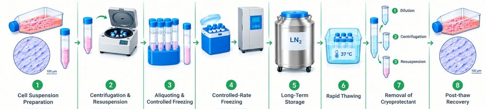
Figure 1: Overview of the Human Dermal Fibroblast Cryopreservation Workflow.
The illustrations in this protocol are provided for indicative purposes only and are intended to support conceptual understanding of the cryopreservation workflow and cellular responses. They are not intended to represent quantitative or cell-specific experimental measurements.
A cooling rate of approximately 1 °C/min has been investigated in fibroblast and keratinocyte cryopreservation systems, although the optimal cooling rate can depend on cell type and cryoprotectant configuration (Whaley et al., 2021).
The specific cell concentration, CPA exposure duration, centrifugation parameters, CPA‑removal procedure, and post‑thaw seeding density should be established and validated for the individual HDF preparation because the cited literature does not define universal values for these parameters.
4. Equipment
-
Cell‑culture incubator maintained under conditions appropriate for mammalian fibroblast culture, including approximately 37 °C and controlled CO₂.
- Centrifuge suitable for recovery of fibroblasts following cell detachment.
- Controlled‑rate freezer or validated gradual‑cooling system capable of providing approximately 1 °C/min cooling.
- −80 °C freezer for the intermediate freezing stage.
- Validated cryogenic storage system for long‑term preservation.
- Validated warming system or water bath maintained at approximately 37 °C .
- Inverted microscope.
- Cell counting and viability assessment system.
- Appropriate biosafety equipment for handling primary human cells.
- Appropriate cryogenic protective equipment.
The cited HDF studies support controlled/gradual cooling and cryogenic storage but do not establish a single universal centrifugation force or cooling‑system configuration. Equipment should therefore be selected according to the validated laboratory workflow.
5. Reagents and Consumables
5.1 Primary Cells
- Primary human dermal fibroblasts derived from human skin.
The starting culture should be actively proliferating and suitable for experimental use. The cited HDF literature does not establish one universal passage number or confluence threshold for cryopreservation; Therefore, these parameters should be documented and defined according to the validated laboratory culture system.
5.2 Principal Cryoprotective Formulation
This formulation has direct experimental support in human fibroblast cryopreservation (Mohamed et al., 2024).
5.3 Alternative Literature‑Supported Formulation
HES -containing, low- DMSO formulation
- HES : investigated as a non-penetrating/macromolecular cryoprotectant
- DMSO : low- DMSO formulations have been investigated
HES ‑containing cryoprotective formulations have been investigated for dermal fibroblast cryopreservation (Whaley et al., 2021).
This formulation should be treated as a separate experimental condition and should not be combined with the principal 10% DMSO formulation.
5.4 Culture Medium
- Fibroblast‑compatible basal culture medium for pre‑freezing culture and post‑thaw recovery.
5.5 Cryogenic Vessels
- Sterile cryogenic vials suitable for the intended storage temperature.
- Appropriate vial identification and labeling materials.
6. Experimental Protocol
6.1 Preparation of the Fibroblast Culture
- Maintain primary HDFs under appropriate adherent culture conditions.
- Use an actively proliferating culture as the starting material.
- Record the cell source, passage number, culture conditions, and date of cryopreservation.
- Avoid cultures showing evident abnormal morphology or poor growth.
- Confirm that the culture is suitable for cryopreservation before initiating cell harvesting.
The available HDF literature does not establish a universal passage number or confluence threshold applicable to all primary HDF preparations. These parameters should therefore be defined according to the validated laboratory culture system.
6.2 Cell Harvesting
- Remove the culture medium from the fibroblast culture.
- Detach the adherent fibroblasts using an appropriate validated enzymatic dissociation procedure.
- Recover the cells as a uniform cell suspension.
- Determine cell concentration.
- Determine pre‑freezing viability using a validated viability assay.
- Record the pre‑freezing cell concentration and viability before CPA exposure.
The cell suspension should provide a defined and reproducible starting population for cryoprotectant exposure.
Critical Control Point
Do not proceed without documenting the starting cell concentration and viability. The cited literature supports assessment of cell viability before cryopreservation but does not establish a universal minimum numerical viability threshold applicable to all primary HDF preparations.
7. Preparation of Cryoprotective Medium
7.1 Principal Formulation
Prepare the cryoprotective medium as:
Nota bene: For the principal formulation, the target is 10% DMSO (v/v) in the final cell-containing suspension. Account for the volume of the harvested cell suspension, including residual culture medium, when calculating the required DMSO and FBS volumes. The cell suspension and FBS together constitute the remaining non-DMSO fraction of the final formulation.
This serum/ DMSO configuration has been used in human fibroblast cryopreservation (Mohamed et al., 2024).
The cryoprotective medium should be prepared under appropriate sterile conditions and handled in a manner that minimizes unnecessary DMSO exposure before freezing.
7.2 Alternative Formulation
A recent alternative formulation approach is:
FBS + 2% DMSO + 0.25% HES (v/v)
HES -containing, low- DMSO formulations have been investigated for human dermal fibroblasts (Zheng et al., 2024).
It should be treated as a separate cryopreservation condition rather than as a modification of the 10% DMSO formulation.
Critical Control Point
DMSO exposure should be controlled because its cytotoxicity is dependent on concentration, temperature, and exposure duration (Weng & Beauchesne, 2020).
The cited HDF literature does not establish one universal DMSO exposure duration applicable to all HDF preparations. The exposure interval should therefore be defined by the validated laboratory procedure.
8. CPA Exposure and Cell Dispensing
- Prepare the fibroblast suspension for addition of the selected cryoprotective medium.
- Add the cryoprotective formulation using a controlled procedure that minimizes localized exposure to concentrated DMSO.
- Mix gently to obtain a homogeneous cell suspension.
- Maintain the CPA exposure for the validated processing interval.
- Dispense the cell suspension into sterile cryogenic vials .
- Use a consistent volume per vial. Approximately 1 mL per cryovial has been used in published human fibroblast cryopreservation procedures.
- Label each vial with:
- cell identity;
- passage number;
- cryoprotective formulation;
- date of freezing;
- relevant sample identifier.
Human fibroblast cryopreservation studies support the use of DMSO‑containing cryoprotective formulations, but the precise exposure interval should be considered formulation‑and workflow‑dependent (Mohamed et al., 2024; Whaley et al., 2021).
Critical Control Point
The interval between CPA addition and initiation of cooling should be controlled and reproducible between samples because prolonged exposure to DMSO can contribute to CPA‑associated toxicity (Weng & Beauchesne, 2020).
Because the cited studies do not establish a universal HDF‑specific CPA exposure time, the validated laboratory exposure interval should be documented in the batch record.
9. Controlled Cooling
9.1 Cooling Rate
Apply controlled or gradual cooling at approximately:
1 °C/min
Controlled cooling at approximately this rate has been investigated in fibroblast and keratinocyte cryopreservation systems (Whaley et al., 2021).
The rationale is to permit sufficient cellular water loss during freezing while reducing the probability of intracellular ice formation, although cryopreservation response remains dependent on cell type and experimental configuration (Whaley et al., 2021).
9.2 Cooling Procedure
- Place sealed cryogenic vials in the validated cooling system.
- Initiate gradual/controlled cooling.
- Maintain a cooling rate of approximately 1°C/min .
- Continue cooling until approximately −80 °C is reached.
Transfer the frozen vials from the intermediate freezing stage to the validated cryogenic storage system.
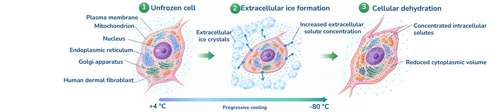
Figure 2: Cellular Responses During Cryopreservation of Human Dermal Fibroblasts.
The illustrations in this protocol are provided for indicative purposes only and are intended to support conceptual understanding of the cryopreservation workflow and cellular responses. They are not intended to represent quantitative or cell-specific experimental measurements.
Critical Control Point
The cooling profile should be monitored or otherwise validated to confirm that the intended cooling rate is achieved.
A cooling rate of approximately 1 °C/min should not be interpreted as a universal optimum for every fibroblast population. Comparative work demonstrates that cooling rate can influence fibroblast and keratinocyte recovery (Whaley et al., 2021).
10. Cryogenic Storage
- Following the initial controlled freezing stage, transfer the frozen vials to a validated cryogenic storage system.
- Maintain samples under conditions appropriate for long‑term cryogenic preservation.
- Record the storage location and date.
- Minimize unnecessary warming and repeated temperature fluctuations during storage.
The −80 °C stage should be considered an intermediate freezing stage rather than the long‑term storage condition in this protocol.
Cryopreservation studies in human primary cells have evaluated the effects of storage duration and revival procedures on post‑thaw recovery (Mohamed et al., 2024).
Under the specific experimental conditions reported by Mohamed et al. (2024), HDFs preserved using an FBS + 10% DMSO formulation showed viability above 80% following evaluated storage periods including 1 and 3 months.
This observation is study‑specific and should not be interpreted as a universal viability guarantee for all HDF populations, cryoprotective formulations, or storage systems (Mohamed et al., 2024).
11. Warming and Post‑Thaw Recovery
11.1 Rapid Warming
- Remove one cryovial from cryogenic storage.
- Warm the sample rapidly using a validated warming procedure at approximately 37 °C .
- Minimize the duration of exposure to partially frozen conditions.
- Once adequately warmed, proceed immediately with dilution and recovery.
The approximately 37 °C value refers to the warming temperature , not a warming rate.
Rapid warming is consistent with established cryobiological principles because prolonged exposure to partially frozen temperature ranges can promote damaging ice growth and recrystallization (Whaley et al., 2021).
12. CPA Dilution and Removal
- Transfer the warmed cell suspension into an appropriate volume of fibroblast‑compatible recovery medium.
- Dilute the suspension to reduce continued exposure to DMSO.
- Apply the validated CPA‑removal procedure for the HDF preparation.
- If centrifugation is part of the validated recovery procedure, recover the cells and resuspend them in fresh culture medium.
Return the cells to appropriate adherent culture conditions.
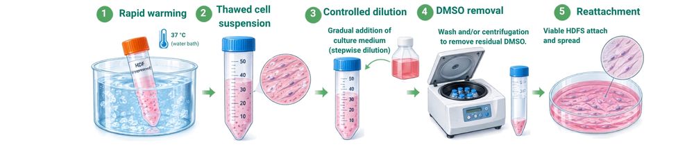
Figure 3: Post-Thaw Recovery of Human Dermal Fibroblasts.
The illustrations in this protocol are provided for indicative purposes only and are intended to support conceptual understanding of the cryopreservation workflow and cellular responses. They are not intended to represent quantitative or cell-specific experimental measurements.
The literature supports more than one post‑thaw recovery strategy and does not establish one universal HDF‑specific CPA‑removal procedure. Consequently, the dilution ratio, centrifugation conditions, and recovery sequence should be validated for the selected HDF population.
Critical Control Point
CPA removal should be sufficiently rapid to limit continued DMSO exposure while avoiding excessive osmotic stress during dilution. DMSO toxicity and osmotic effects are important components of cryoprotectant‑associated cellular injury (Weng & Beauchesne, 2020).
13. Post-Thaw Culture
- Transfer recovered HDFs to appropriate adherent culture conditions.
- Allow the cells to recover and attach.
- Examine cell morphology microscopically.
- Assess post-thaw viability.
- Evaluate cell attachment after approximately 24 hours .
- Continue monitoring cell number and morphology during subsequent recovery.
- Where required, assess proliferation and fibroblast‑associated phenotype.
Immediate post‑thaw viability should not be considered the sole indicator of successful cryopreservation because membrane integrity does not necessarily reflect complete functional recovery.
Assessment of attachment, morphology, and subsequent proliferation can provide additional information about post‑thaw cellular recovery.
14. Quality Control Measures
14.1 Pre-Freezing Quality Control
Record:
- Cell identity.
- Passage number.
- Cultural condition.
- Cell concentration.
- Pre-freezing viability.
- Date of cryopreservation.
- Cryoprotective formulation.
- Relevant sample or batch identifier.
Where appropriate, the culture should also be monitored for contamination, including mycoplasma , before cryopreservation.
14.2 Immediate Post‑Thaw Viability
Assess membrane integrity using a validated viability assay such as dye exclusion or another appropriate method.
Record:
- Total cell count.
- Viable cell count.
- Percentage viability.
Immediate viability provides an important recovery endpoint but does not fully characterize functional recovery.
14.3 Post-Thaw Attachment
Evaluate cell attachment after approximately 24 hours .
Attachment provides a functional recovery endpoint that is not completely captured by immediate membrane integrity measurements.
14.4 Morphology
Assess:
- Cell attachment.
- Fibroblast morphology.
- Evidence of abnormal cellular appearance.
- Recovery of the expected adherent phenotype.
14.5 Proliferation
Where required, evaluate subsequent cell proliferation.
Ki‑67 should be interpreted as a proliferation‑associated marker rather than a fibroblast identity marker.
Where Ki‑67 is used, it should therefore be reported as an endpoint reflecting proliferative activity rather than fibroblast identity.
14.6 Fibroblast-Associated Phenotype
Where phenotype preservation is important, fibroblast‑associated markers such as collagen‑I may be evaluated.
The selected markers should be appropriate to the specific biological question and should not be interpreted as universal identifiers of all human fibroblast populations.
14.7 Cellular Structures
Assessment of nuclei, mitochondria, actin organization, or other relevant cellular structures can provide information beyond simple membrane integrity measurements.
15. Acceptance and Interpretation Criteria
The available peer‑reviewed HDF literature does not establish a single universal numerical acceptance criterion for post‑thaw viability, attachment, proliferation, or phenotype.
Therefore, cryopreservation performance should be interpreted using multiple complementary endpoints.
Successful Recovery
A favorable recovery profile is characterized by:
High post‑thaw viability + appropriate attachment + expected morphology + continued proliferation
This combination provides stronger evidence of successful HDF recovery than immediate viability alone.
High Viability but Poor Attachment
High immediate viability accompanied by poor attachment may indicate sublethal cryoinjury that is not detected by membrane integrity assays alone.
Potentially affected processes include:
- Cell adhesion.
- Cytoskeletal organization.
- Mitochondrial function.
- Cellular recovery.
Low Post-Thaw Viability
Potential contributing mechanisms include:
- Inadequate cryoprotection.
- Excessive DMSO toxicity.
- Inappropriate cooling.
- Excessive solution‑effect injury.
- Intracellular ice formation.
- Warming-associated injury.
A specific mechanism should not be assigned solely from viability measurements because several cryoinjury mechanisms can produce similar post‑thaw phenotypes (Whaley et al., 2021; Weng & Beauchesne, 2020).
Preserved Viability but Abnormal Morphology
This may indicate incomplete functional recovery or structural injury despite preserved membrane integrity.
Differences Between Cryoprotective Formulations
Results obtained with FBS + 10% DMSO should not automatically be extrapolated to HES -containing, low-DMSO formulations , or vice versa.
The formulations differ in CPA composition and have been evaluated under different experimental configurations (Mohamed et al., 2024; Zheng et al., 2024).
Differences Between Suspension and Adherent Freezing
Cryopreservation outcomes can differ according to whether fibroblasts are frozen as a cell suspension or under an adherent configuration. Results obtained using one configuration should therefore not automatically be extrapolated to another.
16. Critical Parameters Requiring Laboratory Validation
The following parameters should be defined and documented in the laboratory's validated procedure because the cited HDF literature does not establish universal values:
| parameter | Recommended approach |
|---|---|
| Passage number | Record and validate according to the HDF culture system |
| Pre-freezing confluence | Define during laboratory validation |
| Starting viability | Establish a laboratory‑specific acceptance criterion |
| Cell concentration | Validate for the selected HDF population |
| CPA addition procedure | Standardize and document |
| CPA exposure duration | Validate because DMSO toxicity is exposure‑dependent |
| Centrifugation force/time | Validate for the selected recovery procedure |
| CPA dilution ratio | Validate for osmotic compatibility |
| Post-thaw seeding density | Define according to the recovery system |
| Cooling system | Validate the selected controlled/gradual cooling method |
| Cooling rate | Approximately 1 °C/min as a literature‑supported starting condition |
| Intermediate freezing stage | Approximately −80 °C |
| Warming temperature | Approximately 37 °C |
| Long-term storage | Validated cryogenic storage conditions |
| Post-thaw assessment | Viability, attachment, morphology and, where relevant, proliferation/phenotype |
This distinction is intentional: parameters supported directly by the cited HDF literature are stated explicitly, whereas parameters not established as universal are identified for laboratory validation rather than assigned arbitrary values.
17. Troubleshooting
| Observation | Possible explanation |
|---|---|
| Low immediate post‑thaw viability | CPA toxicity, inadequate cryoprotection, inappropriate cooling, intracellular ice formation, solution‑effect injury, or warming‑associated injury |
| High viability but poor attachment | Sublethal injury affecting adhesion, cytoskeleton, mitochondria, or cellular recovery |
| Poor morphology after thawing | Cellular or structural cryoinjury |
| Good immediate viability but poor proliferation | Sublethal injury not detected by membrane integrity assays |
| Different outcomes between formulations | Differences in CPA composition and cryopreservation configuration |
| Different outcomes between suspension and adherent freezing | Configuration‑dependent cryoinjury and recovery |
Troubleshooting should consider the complete cryopreservation sequence rather than attributing an outcome to viability alone.
18. Conclusion
Cryopreservation of primary human dermal fibroblasts enables viable cell populations to be preserved for subsequent research. A DMSO‑containing cryoprotective formulation combined with controlled/gradual cooling, cryogenic storage, rapid warming, controlled CPA dilution, and multidimensional post‑thaw assessment provides a literature‑supported strategy for preserving primary HDF populations (Mohamed et al., 2024; Whaley et al., 2021).
The principal formulation described in this protocol is 90% FBS + 10% DMSO , while HES- containing, low-DMSO formulations represent an alternative formulation approach. These conditions should be evaluated independently because cryopreservation outcome depends on CPA composition, cellular configuration, cooling conditions, storage, and recovery procedures (Zheng et al., 2024; Mohamed et al., 2024).
For reproducible implementation, laboratory‑specific parameters that are not established as universal in the HDF literature—particularly cell concentration, CPA exposure interval, centrifugation conditions, CPA dilution procedure, and post‑thaw seeding conditions—should be validated and documented before routine use.
References
- Weng L, Beauchesne PR. (2020). Dimethyl sulfoxide-free cryopreservation for cell therapy: A review. Cryobiology , 94:9–17 . doi:10.1016/j.cryobiol.2020.03.012.
- Whaley D, Damyar K, Witek RP, Mendoza A, Alexander M, Lakey JRT. (2021). Cryopreservation: An Overview of Principles and Cell-Specific Considerations. Cell Transplantation , 30 . doi:10.1177/0963689721999617.
- Murray KA, Gibson MI. (2022). Chemical approaches to cryopreservation. Nature Reviews Chemistry , 6:579–593 . doi:10.1038/s41570-022-00407-4.
- Kardorff M, Mahler HC, Huwyler J, Jere D, Sorret L (2023). Cryoprotection in Human Mesenchymal Stromal/Stem Cells: Synergistic Impact of Urea and Glucose. Journal of Pharmaceutical Sciences , 112(6):1681–1686 . doi:10.1016/j.xphs.2023.02.001.
- Martín-López M, Rosell-Valle C, Arribas-Arribas B, et al. (2023). Bioengineered tissue and cell therapy products are efficiently cryopreserved with pathogen-inactivated human platelet lysate-based solutions. Stem Cell Research & Therapy , 14:69 . doi:10.1186/s13287-023-03300-z.
- Mohamed HM, Sundar P, Ridwan NAA, et al. (2024). Optimization of cryopreservation conditions, including storage duration and revival methods, for the viability of human primary cells. BMC Molecular and Cell Biology , 25(1):20 . doi:10.1186/s12860-024-00516-6.
- Zheng M, Hu T, Yang Y, et al. (2024). In situ-formed cryomicroneedles for intradermal cell delivery. NPG Asia Materials , 16:11 . doi:10.1038/s41427-024-00531-1.